|
|
Soft X-Ray Tomography in MST Paolo Franz
|
|
|
Soft X-Ray Tomography in MST Paolo Franz
|
| Contents Description Electronic layout User manual Manuale Info Images Results History Logbooks Bibliography Links |
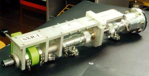
See the complete history clicking here. This web page summarises and describes the latest ideas and results about the diagnostic for measuring soft x-ray (SXR) profiles in MST [Dexter R N, et al., Fusion Technol. 19, 131 (1991)] we designed and entirely built, installed in MST between November 2001 and August 2004. All these activities are included in the existing collaboration between the RFX [Rostagni G, Fusion Engineering and Design 25, 301 (1995)] and MST group dedicated to the project, the development and the use of a novel soft x-ray tomographic system for imaging of MHD phenomena in the Reversed Field Pinch (RFP).
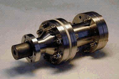
The rich variety of MHD phenomena in the RFP calls for advanced, non-invasive imaging systems that allow the diagnostic of the plasmas with high spatial and temporal resolution. This is the case, for example, to follow the non-linear evolution of MHD instabilities, in particular in those regimes where they are observed to drastically change their properties. This happens, for example, as a result of active fluctuations control techniques like poloidal current drive or in self-organised laminar states as those obtained in the Quasi Single Helicity (QSH) regime. SXR imaging of the plasma is a very powerful technique for this purpose. A neat example has been provided by the RFX group, which has developed a powerful SXR tomography for the RFX device [Martin P, et al., Rev. Sci. Instrum. 68, 1256 (1997), Franz P, et al., Nucl. Fusion 41, 695 (2001)]. This diagnostic enabled the achievement of a large number of new results, including the finding of QSH states. Researchers of the RFX group developed a flexible, new, miniaturised SXR camera that could be used to monitor not only the slow evolution of the SXR emissivity pattern, but also its high frequency fluctuations. Given its small size and flexible design, this camera is portable to many different plasma devices. The MST RFP device represented a unique opportunity for this development. On the one hand it offers a flexible and easy-to-use experimental RFP facility, with minor radius equal to that of RFX. On the other side the use of a SXR imaging system in this device has provided a significant amount of new information on MHD dynamics in an experiment with smooth boundary conditions. Information on plasma structure and MHD dynamics in a variety of conditions, including advanced confinement regimes and QSH states are extremely useful for the understanding of RFP physics and to gain experience for the future RFP operation. This web-site focuses mainly on the SXR photocameras (probes), which have been inserted in the 1.5 inch portholes of a particular poloidal section of the MST vacuum vessel, which form the tomographic system. A few papers describing the diagnostic have been published (P.Franz, et al., Rev. Sci. Instrum. 74, 2152 (2003), P.Franz, et al., Rev. Sci. Instrum. 75, 4013 (2004) and P.Franz, et al., Rev. Sci. Instrum. 77, 10F318 (2006)). Physical results can be found for example on P.Franz, et al., Physics Review Letters 92, 125001 (2004) or P.Franz, et al., Physics of Plasmas, 13, 012510 (2006); see also the bibliography page. Scientists of Consorzio RFX involved in the collaboration: |
| Description | Electronic layout | User manual | Manuale | Info | Images | Results | History | Logbooks | Bibliography | Links |
| Copyright © 2001-2013 Paolo F Version 1.1 This page was last modified: |